
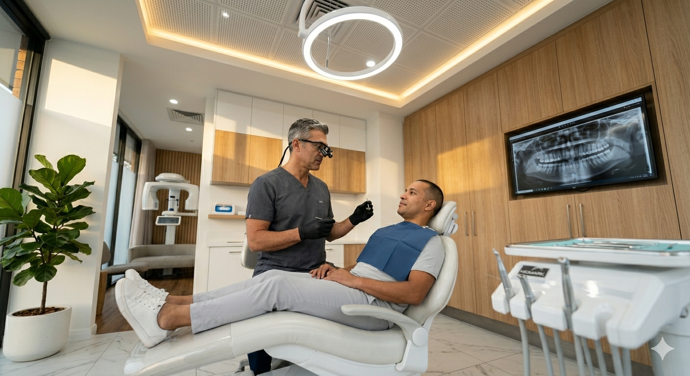
You will discover how modern implant dentistry in Oregon combines proven science with patient-centered care to restore smiles and chewing comfort. Cutting-edge imaging, advanced materials, and personalized planning let you get implants that look natural and feel secure.
A neighborhood dental expert explains how implants work, what modern innovations change about the experience, and how safety and comfort guide every step. This article breaks down techniques, choices in materials, and practical next steps so you can decide confidently about treatment options.
Expect clear explanations of what to ask your dental professional, how recovery typically goes, and how ongoing care preserves results—plus tips on choosing the right team for your smile care.
Dental implants replace missing teeth with a stable, long-term solution that preserves jawbone and restores function. Readers will learn what implants are, how they benefit oral health, and who typically qualifies for treatment.
A dental implant is a small titanium screw surgically placed into the jawbone to serve as an artificial tooth root. After osseointegration — the process where bone fuses to titanium — a connector (abutment) and a crown, bridge, or denture attach to the implant to restore chewing and appearance.
Key components:
Implants can replace a single tooth, several teeth, or support full-arch prostheses. Modern planning uses 3D imaging (CBCT) and guided surgery to optimize position and avoid nerves and sinuses. Healing time varies but typically ranges from 3 to 6 months before final restoration.
Dental implants preserve adjacent teeth because they do not require grinding down neighboring teeth like traditional bridges. They maintain jawbone volume by transmitting chewing forces to bone, which reduces the bone loss that follows tooth extraction.
Function and comfort improve: implants restore bite strength similar to natural teeth, enabling normal chewing and clearer speech. They also reduce the risk of shifting teeth and bite collapse that occur with long-term tooth loss. Biocompatible materials lower the risk of allergic reaction, and good oral hygiene plus regular check-ups help implants last many years. Patients often report higher confidence and easier maintenance compared with removable dentures.
Suitable candidates are adults with good overall health, adequate jawbone volume, and healthy gum tissue. Chronic conditions like uncontrolled diabetes or habits such as heavy smoking can impair healing; these factors require evaluation and management before implant placement.
Bone deficiency does not automatically disqualify a patient. Bone grafting, sinus lifts, or zygomatic implants can provide alternatives when native bone is insufficient. Clear steps in assessment include medical history review, dental exam, and 3D imaging to plan implant size and position. The dental team discusses expectations, timeline, costs, and any necessary preparatory treatments so patients can make an informed decision.
These advances streamline diagnosis, improve surgical precision, and reduce recovery time. They also increase predictability of outcomes and help tailor treatment to each patient’s anatomy and comfort needs.
Digital cone-beam CT (CBCT) provides high-resolution, 3D views of the jaw, sinuses, and nerve pathways, letting clinicians measure bone volume and density precisely. In practice, this reduces guesswork when selecting implant size and position.
Intraoral scanners capture accurate digital impressions without messy putty. The resulting STL files integrate with planning software and CAD/CAM mills to produce provisional crowns, custom abutments, and surgical guides faster than traditional lab workflows.
Typical benefits for patients include fewer visits, smaller errors between planned and actual restorations, and clearer visual explanations before treatment. Clinics often pair CBCT with optical scans to create a complete digital model for simulation and patient education.
Computer-guided surgery uses planning software to align virtual implant placement with prosthetic goals. The clinician reviews bone anatomy, prosthetic space, and nerve locations, then finalizes implant depth and angulation in the software.
A 3D-printed or milled surgical guide transfers that plan to the mouth, guiding drills and implants along the exact trajectory. This increases placement accuracy and reduces chair time compared with freehand techniques.
Guided workflows reduce intraoperative surprises, lower the chance of encroaching on critical anatomy, and make immediate provisionalization more predictable. For complex cases—limited bone, angled implants, or full-arch restorations—guided surgery improves consistency and patient confidence.
Minimally invasive implant techniques prioritize tissue preservation and faster healing. Flapless placement, when appropriate, avoids cutting and suturing gum tissue; it uses small punch access and the implant is placed through the existing mucosa.
Shorter surgical time and less postoperative pain typically follow. When bone grafting is needed, socket preservation and ridge augmentation use refined biomaterials and membranes that integrate predictably with native bone.
Immediate-load protocols let certain patients receive a fixed provisional tooth the same day as implant placement. Careful case selection—adequate bone, primary stability above recommended thresholds, and good oral hygiene—is essential for success and reduced complication risk.
This section highlights materials and protocols that improve implant integration, esthetics, and treatment speed. It focuses on biocompatible surfaces, patient-specific components, and protocols that allow safe early function.
Modern implants use titanium alloys and zirconia because tissues tolerate them well and they resist corrosion.
Grade 4 commercially pure titanium and titanium-6Al-4V ELI are common; they combine strength with proven osseointegration. Surface treatments — such as sandblasting, acid etching, and plasma spraying — increase surface roughness to promote faster bone contact and stronger mechanical interlock.
Zirconia implants suit patients with metal sensitivities or high esthetic demands. They are white, reducing gray gum show-through, and offer good soft-tissue response, though long-term data remain more limited than titanium’s.
Clinicians choose material based on bone quality, loading demands, and esthetic zone requirements.
Key considerations:
Custom abutments are CAD/CAM-milled components tailored to the patient’s gingival profile and prosthetic plan.
They can be made from titanium, zirconia, or hybrid materials depending on esthetic needs and soft-tissue thickness.
Custom design allows precise emergence profiles that support papillae and reduce food traps. This improves esthetics and facilitates hygiene.
Clinicians use digital intraoral scans and 3D implant position data to design abutments that match crown contours, minimizing crown adjustments and chair time.
Benefits include:
Immediate load protocols place a restoration on the implant within 48–72 hours of surgery to restore function and appearance quickly.
Success depends on achieving primary stability, typically insertion torque >30–35 Ncm and favorable bone density (D1–D3).
Surgical techniques include under-preparation of the osteotomy or use of tapered, roughened implants to enhance mechanical engagement. Prosthetic measures—such as splinting multiple implants and limiting occlusal contacts—reduce micromovement risk during healing.
Careful case selection and controlled loading are critical; patients with poor bone quality, parafunction, or uncontrolled systemic conditions are less suitable.
Clinical steps for predictable outcomes:
If patients want faster results, clinicians should discuss immediate load risks and alternatives and confirm suitability with diagnostic imaging and systemic health review.
This section explains how clinicians tailor implant treatments using detailed digital records and image-guided planning to match each patient’s anatomy, bite, and cosmetic goals. It highlights tools that improve accuracy, reduce surprises during surgery, and communicate outcomes clearly.
Virtual Smile Design (VSD) uses photographs, intraoral scans, and software to create a photorealistic preview of expected tooth position, shape, and color. The dentist imports the patient’s digital impressions and facial photos, adjusts tooth proportions and midline, and simulates restorative materials so the patient can see likely results before any irreversible work begins.
VSD guides decisions about extraction, bone grafting, and implant position to achieve both function and appearance. It also generates visuals for lab communication and provisional restorations, reducing miscommunication and remakes. Patients appreciate seeing specific changes—veneers, crown heights, or gingival contours—so consent is informed and expectations align with clinical limits.
Cone Beam CT (CBCT) provides 3D images of jawbone volume, nerve location, and sinus anatomy essential for safe implant placement. Scans measure bone height and width in millimeters, reveal cortical thickness, and identify angulation issues that 2D X-rays can miss.
Clinicians import CBCT data into planning software to plan implant length, diameter, and exact entry angle, then design a surgical guide if needed. CBCT also helps assess pathology—cysts, impacted teeth, or bone defects—that changes the surgical approach or indicates grafting. Radiation dose is low compared with medical CT, and modern units use focused fields of view to minimize exposure.
Modern implant dentistry focuses on predictable chewing, clear speech, and a comfortable bite while matching natural tooth appearance. Patients benefit from implant choices that prioritize stable support, soft-tissue health, and prostheses shaped for easy cleaning.
Implant crowns are custom-made from materials like zirconia or layered porcelain to match the patient’s tooth color, translucency, and shape. Technicians use digital scans and shade-matching systems to reproduce adjacent tooth anatomy, so restorations blend seamlessly during speech and smiling.
Soft-tissue contouring around the implant matters as much as the crown. Precision abutments and healing collars shape the gingiva to mimic natural papillae, reducing dark triangles and improving hygiene access.
Esthetic planning often includes mock-ups and try-ins. These allow clinicians to adjust tooth length, midline, and occlusal contacts before finalizing the prosthesis, ensuring both comfort and a lifelike result.
Full-arch implant restorations replace all teeth on an upper or lower jaw using 4–8 implants for fixed bridges or fewer for implant-retained overdentures. Treatment choices depend on bone volume, bite forces, and the patient’s desire for a removable versus fixed prosthesis.
Immediate-load protocols can provide a temporary fixed bridge the day of surgery when primary stability meets clinical thresholds (typically insertion torque ≥30 Ncm). Final prostheses use milled frameworks and layered ceramics or high-density zirconia to resist wear and restore proper vertical dimension.
Maintenance planning includes routine hygiene visits, occlusal checks, and replacement of wear components like nylon housings or screw-retained attachments. Clear aftercare instructions and scheduled follow-ups help patients preserve comfort, function, and long-term success.
They should contact their dental team to review which full-arch option fits their health, budget, and lifestyle.
Patients should expect clear explanations of sedation choices, step-by-step care instructions, and evidence-based infection control to reduce anxiety and promote healing. Communication about risks, costs, and timelines also helps patients make informed decisions and prepares them for recovery.
Clinics typically offer local anesthesia, nitrous oxide (laughing gas), oral sedatives, and IV sedation depending on patient anxiety and procedure complexity. Local anesthesia numbs only the implant site; it is used in most straightforward cases and lets patients stay awake and responsive.
Nitrous oxide provides mild relaxation and rapid recovery, making it a common choice for anxious but cooperative patients. Oral sedatives like diazepam or triazolam are useful for moderate anxiety; patients must arrange a ride home.
IV sedation delivers deeper sedation and is reserved for long surgeries, severe anxiety, or multiple implants in one visit. Monitoring includes pulse oximetry, blood pressure, and heart rate, and a trained clinician must manage the airway. Clinics should document medical history, current medications (especially blood thinners), and allergies before any sedation decision.
Immediate post-op instructions focus on bleeding control, swelling management, pain relief, and infection prevention. Patients should bite gently on gauze for 30–60 minutes to control bleeding, apply ice packs intermittently for the first 24 hours, and keep the head elevated when resting.
Prescribed analgesics (acetaminophen or NSAID combinations) and, when indicated, short antibiotic courses reduce discomfort and infection risk; clinicians tailor prescriptions to patient health status. Soft foods and avoiding straw use help protect the surgical site during the first 7–10 days.
Oral hygiene must continue with gentle rinsing starting 24 hours after surgery, using saline or a chlorhexidine rinse if recommended. Follow-up visits at 1–2 weeks confirm initial healing, and imaging (periapical X-ray or CBCT) verifies implant position before loading with a crown, typically after osseointegration of 3–6 months.
Implant maintenance and new technologies both aim to keep restorations functional, comfortable, and long-lasting. Practical daily care, regular professional checks, and emerging digital tools are the pillars patients should expect.
They should follow a structured home-care routine: soft-bristled toothbrush twice daily, interdental brushes sized to the space, and daily use of low-abrasive fluoride toothpaste. Flossing under bridges or around implant crowns needs either waxed floss, floss threaders, or water flossers to remove plaque without damaging the prosthetic margins.
Clinics typically schedule professional follow-ups every 3–6 months during the first year, then at least biannually. At visits, the clinician will check implant stability, peri-implant tissue health, occlusion, and prosthetic fit; they may take radiographs to monitor bone level. If signs of inflammation or bone loss appear, early interventions include focused cleaning, local antiseptics, and guided antimicrobial therapy to prevent progression.
Patients with bruxism, smoking history, diabetes, or poor oral hygiene require more frequent monitoring and possibly occlusal guards or lifestyle counseling. Clear, written home-care instructions and demonstrations improve compliance and reduce complications.
Digital dentistry continues to refine implant workflows: intraoral scanners create precise digital impressions, and CAD/CAM milling or 3D printing produces custom abutments and restorations with tighter tolerances. This reduces lab turnaround and improves fit, which lowers mechanical complications like loosening or fracture.
Artificial intelligence assists in treatment planning by analyzing CBCT scans to suggest optimal implant size, position, and angulation based on anatomical landmarks and prosthetic needs. Guided surgery using fully digital guides or dynamic navigation increases placement accuracy and reduces chair time in complex cases.
Emerging surface coatings and biomaterials aim to enhance osseointegration and resist bacterial colonization. Also, remote monitoring tools and patient apps can track symptoms, prompt hygiene actions, and flag issues for early clinic review. Patients should ask their clinician how these technologies apply to their individual care plan.
Patients should look for a dentist or specialist with specific implant training and current credentials. Many general dentists place implants, but periodontists and oral surgeons often handle more complex surgical cases; prosthodontists focus on the restorative, cosmetic outcome.
Ask about experience: number of implant cases per year, types of systems used, and complication rates. Request before-and-after photos and patient references to understand typical results and patient comfort.
Verify credentials and memberships. Check state licensing, board certifications, and memberships in organizations like the American Academy of Implant Dentistry or International Congress of Oral Implantologists for continuing education.
Consider the practice environment and patient experience. Look for clear communication, digital planning tools (CBCT, intraoral scanning), sedation options, and a care team that explains each step calmly and honestly.
Use this checklist when comparing clinicians:
Encourage a consultation visit to assess rapport and comfort. A brief meeting lets patients ask about timelines, warranties, and what to expect during recovery.
Invite them to bring questions and records to that consultation to make a confident, informed decision about implant care.
They should schedule an initial consultation to review medical history, oral health, and cosmetic goals. This visit lets the implant dentist explain options, timelines, and likely outcomes in plain language.
A clear treatment plan follows, often including imaging like CBCT scans and a discussion of anesthesia or sedation choices. The plan will outline costs, insurance considerations, and any preparatory treatments such as extractions or bone grafting.
Daily home care remains essential before and after implant placement. Brushing twice daily, flossing, and using an interdental brush reduce infection risk and support long-term success.
Regular professional checkups and cleanings help the clinician monitor healing and spot early issues. Typical follow-up visits occur several times in the first year, then shift to routine maintenance every six months.
They can ask about restorative options for final crowns, bridges, or overdentures to match natural teeth in color and function. The dentist will explain materials, durability, and expected lifespan so patients can make informed choices.
If anxiety or pain is a concern, discuss sedation and pain-control strategies ahead of the procedure. Many practices offer nitrous oxide, oral sedation, or local anesthesia tailored to comfort and safety.
To move forward, contact the dental office to book a consultation or request pre-visit instructions. Gentle guidance and clear answers help patients feel prepared and confident about improving their smile.
This section answers practical questions about 2026 implant advances, less invasive placement, digital planning, smart implants, autoimmune considerations, and how to pick the right clinician. Each item gives clear, patient-focused details that help readers weigh comfort, healing, and long-term outcomes.
Titanium and zirconia implants now use surface coatings that speed bone integration by promoting cell attachment and reducing early inflammation. Shorter healing times—often weeks rather than months—are reported in studies when those surfaces are combined with immediate-loading protocols.
New analgesic and anti-inflammatory delivery systems, including localized drug-eluting collars, help control postoperative pain and reduce swelling. These reduce reliance on systemic pain meds and improve early comfort for most patients.
Flapless surgery with guided osteotomy cuts tissue manipulation and bleeding compared with traditional flap approaches. When bone volume is adequate, dentists often use fully guided drills that limit drilling depth and diameter to what’s needed.
In selected cases, mini-implants and short implants avoid extensive bone grafting and can eliminate wide osteotomies. Some centers use piezoelectric bone surgery to cut bone with less heat and soft-tissue risk than rotary drills.
Cone beam CT scans give three-dimensional views of bone, nerves, and sinus anatomy so clinicians plan implant size and position precisely. Digital implant planning software simulates implant placement and prosthetic outcomes before any surgery occurs.
Guided surgical templates translate the plan to the mouth, reducing human error and improving implant angulation and depth control. This workflow shortens chair time and increases predictability for immediate or delayed restoration.
Smart implants incorporate sensors that measure temperature, load, or micro-movements around the implant-abutment interface. Data transmitted to a clinician’s device can signal early inflammation, overload, or loosening before symptoms appear.
Early clinical use focuses on postoperative monitoring to tailor follow-up and intervene sooner if problems arise. Regulatory approval and long-term outcome data are still expanding, so adoption varies by practice.
Many patients with well-controlled autoimmune diseases—like rheumatoid arthritis or lupus—receive implants successfully when systemic disease activity is low. Preoperative medical clearance and coordination with the patient’s rheumatologist reduce risks related to immunosuppressive medications.
Dentists may adjust timing of biologic or immunomodulatory drugs, use prophylactic antibiotics selectively, and plan longer healing windows. Close postoperative monitoring for infection and delayed osseointegration is standard.
A prosthodontist specializes in replacing teeth and complex restorations; they are a strong choice for full-arch work, complicated esthetic cases, or when multiple disciplines must coordinate. General dentists with advanced implant training can manage single implants and straightforward restorations effectively.
Ask about the clinician’s implant training, number of cases performed annually, complication management, and whether they work with a surgical or restorative specialist when needed. Seek examples of recent before-and-after cases and patient reviews, and request a clear treatment plan with risks, alternatives, and timelines.